
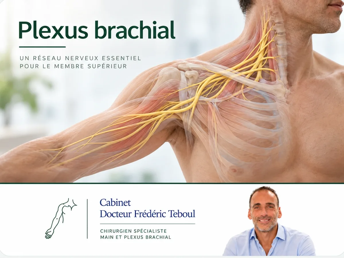
The page devoted to the brachial plexus explains what this condition is, its causes and its work-up. This page goes further: it describes how an injured plexus is reconstructed, what logic guides the surgical choices, and why this surgery is a distinct subspecialty in its own right.
A paradigm shift: from graft to transfer
For a long time, reconstructing a plexus mainly meant grafting — bridging the gap between two nerve endings with nerve segments harvested elsewhere, from the injured root all the way to its target. This “proximal” approach remains useful, but it has a limitation: the distance to cover is long, regrowth is slow, and the target muscle can deteriorate before the fibers reach it.
The turning point of recent decades: selective distal neurotization
Rather than rebuilding the nerve from its origin, a functional donor nerve close to the target is connected directly to the nerve to be reactivated. A 2026 review describes this shift “from proximal reconstruction toward selective distal neurotization strategies.”
The advantage is twofold: regrowth is shorter, and therefore faster, and the function to be restored is targeted precisely. It is this shift that has transformed the outlook for these injuries.
The logic of prioritization: restoring what matters most first
The principle that governs the entire strategy is that not everything can be restored, so priorities must be set. The available nerve resources — the donor nerves — are limited; they are assigned first to the functions on which independence depends most.
Elbow flexion first: without it, the hand cannot be brought to the mouth or the body. It is the priority target of most reconstructions.
Shoulder stability and abduction-rotation, to position the limb in space.
Protective sensation in the hand, to avoid repeated injuries.
And finally control of the hand, the most difficult to restore and often the most incomplete.
A nerve transfer — or neurotization — means sacrificing a secondary nerve function in order to restore an essential one. A donor nerve whose loss is well tolerated is harvested and connected to the paralyzed nerve, close to the target muscle. The brain then relearns how to control the new function. Several transfers have become standard references.
For elbow flexion
One of the most emblematic transfers involves harvesting a few fascicles of the ulnar nerve — without compromising hand function — and connecting them to the nerve of the biceps muscle, restoring elbow flexion. Dr Teboul is co-author of several of the foundational papers describing this technique within the school that developed it.
Teboul F et al. “Transfer of some fascicles from the ulnar nerve to the nerve to the biceps muscle.” Techniques in Hand and Upper Extremity Surgery, 2002; and Teboul F et al. “Transfer of fascicles from the ulnar nerve to the nerve to the biceps in the treatment of upper brachial plexus palsy.” Journal of Bone and Joint Surgery Am, 2004.
The early date of these publications, among the first on nerve transfers, reflects Dr Teboul's active role in the development of nerve and brachial plexus surgery.
In cases of complete brachial plexus palsy with avulsion of all the roots, two options remain in nerve microsurgery: transfer of the intercostal nerves to the motor branch of the musculocutaneous nerve, to restore elbow flexion by reinnervating the biceps and the brachialis muscle; or, as a last resort when the intercostal nerves are contraindicated, a graft between the external branch of the spinal accessory nerve and the motor branch of the musculocutaneous nerve. Finally, a free neurovascular muscle transfer can be performed: harvesting a muscle from the thigh with its nerve and vessels and grafting it to the arm by performing the nerve and vascular anastomoses, in order to create a new muscle to restore elbow flexion as the priority.
For the shoulder: a technique described by Dr Teboul's team
Restoring shoulder function — in particular external rotation, essential for positioning the arm — is a recognized challenge. The usual donors (spinal accessory nerve, partial root) are not always available or sufficient.
An original contribution to the field
Dr Teboul and one of his colleagues described and developed the transfer of the nerve to the rhomboid muscle (a branch of the dorsal scapular nerve) to the suprascapular nerve, performed through a posterior approach as close as possible to the supraspinatus muscle — which shortens the reinnervation pathway.
Anatomical feasibility study: Chirurgie de la Main, 2015. First clinical report: Goubier JN, Teboul F. Hand Surgery & Rehabilitation, 2016. Results on shoulder external rotation: Injury, 2020.
This technique, now cited in the leading international journals on the plexus, illustrates a practice that does not merely apply existing procedures but helps create new ones. However, because of anatomical variations, this nerve transfer is difficult to reproduce and rarely performed. It can be useful in a few very specific cases.
For avulsed roots: extraplexal donors
When several roots are torn from the spinal cord, the donors located within the plexus are lacking. Donors outside the plexus are then used — the spinal accessory nerve, the intercostal nerves, sometimes a controlled portion of another root — to reintroduce control into the limb. The choice and combination of these donors require careful planning, specific to each patient.
The time factor, a defining constraint on strategy
This entire strategy is subject to an unavoidable biological constraint: a muscle deprived of its nerve deteriorates over time. Beyond a certain point, even a technically successful transfer no longer finds a muscle able to respond.
This is what makes plexus surgery as much about the right timing as the right technique. The useful window for reinnervation is measured in months, and the planning — clinical work-up, imaging, electromyography, decision — must fit within that timeframe. Meta-analyses on the restoration of elbow flexion highlight the unfavorable effect of surgical delay on outcomes. Early referral to a specialized center is therefore not a convenience: it determines the result.
Secondary reconstruction: when the nerve is no longer enough
When the window has passed, or as a complement to a nerve reconstruction, palliative techniques that no longer rely on the nerve are used: tendon and muscle transfers, which redirect a healthy muscle to provide a lost function, and sometimes stabilizing fusions. These techniques broaden the range of solutions beyond the nerve window alone, and are an integral part of the plexus surgeon's toolkit.
Team-based, long-term care
Surgery is only one step in a long journey. Specialized rehabilitation supports reinnervation over many months, motor retraining helps the brain take ownership of the newly transferred functions, and the management of neuropathic pain is built in from the outset. The quality of the result depends as much on this continuity as on the surgical procedure itself.
Summary points
These points sum up the logic of the expertise: the right transfer, for the right function, at the right time.
Frequently asked questions
Glossary
Connecting a functional donor nerve to a paralyzed nerve, near the target muscle.
Nerve of the posterior rotator cuff; restoring it recovers part of shoulder function.
A branch of the dorsal scapular nerve, usable as a donor for the suprascapular nerve.
A nerve located outside the plexus (spinal accessory, intercostal) used when local donors are lacking.
Tendon or muscle transfers performed when the window for nerve repair has passed.
Scientific references
A selection of works underpinning the expertise presented. Publications authored by Dr Teboul are shown in bold.
Teboul F et al. Transfer of the rhomboid nerve to the suprascapular nerve: an anatomical feasibility study. Chirurgie de la Main, 2015.
Teboul F et al. Rhomboid nerve transfer to the suprascapular nerve for shoulder reanimation in brachial plexus palsy: a clinical report. Hand Surgery & Rehabilitation, 2016.
Teboul F et al. Transfer of the rhomboid nerve for restoration of shoulder external rotation in partial brachial plexus palsy. Injury, 2020.
Teboul F et al. Transfer of some fascicles from the ulnar nerve to the nerve to the biceps muscle. Techniques in Hand and Upper Extremity Surgery, 2002.
Teboul F et al. Transfer of fascicles from the ulnar nerve to the nerve to the biceps in the treatment of upper brachial plexus palsy. Journal of Bone and Joint Surgery (Am), 2004.
Motor Nerve Transfers in Complete and Incomplete Brachial Plexus Injuries: A State-of-the-Art Review. Neurology International, 2026 — review citing the rhomboid nerve transfer technique.
The Effectiveness of Different Nerve Transfers in the Restoration of Elbow Flexion in Adults Following Brachial Plexus Injury: A Systematic Review and Meta-Analysis. Journal of Hand Surgery, 2023 — effect of surgical delay on outcomes.
Page reviewed and approved by Dr Frédéric Teboul, hand, brachial plexus and peripheral nerve surgeon, member of the Académie Nationale de Chirurgie, co-founder of the Institut de chirurgie du plexus brachial et des nerfs périphériques (Paris).
Last updated: August 20, 2026
Disclaimer. This page is intended to provide information about a specialized practice. It does not replace a consultation. Upper-limb palsy after trauma warrants early specialized referral.
Author of this publication
Every piece of content is signed by its author and reviewed before publication.
 Author
Dr Frédéric Teboul
Brachial plexus and peripheral nerve surgeon
Author
Dr Frédéric Teboul
Brachial plexus and peripheral nerve surgeon