
A lump has appeared on your finger or your wrist. It may not hurt, but it is there, and the first question that comes to mind is never said out loud.
We may as well answer it right away, plainly and without over-reassurance: the vast majority of these masses are benign, and many need no treatment at all. Malignant lesions do exist, they are rare, and they have identifiable features — which is precisely why a persistent mass deserves to be examined rather than watched blindly.
Signs that warrant a prompt opinion
This section comes first because it is the one most useful to you. None of these signs means the lesion is serious. Each one means it deserves to be looked at by a specialist rather than left under observation.
What should prompt a visit
—A mass that keeps growing steadily over weeks or months.
—A hard mass that does not move under the fingers and seems fixed to the deep tissues.
—A mass larger than two centimeters, or lying deep rather than just under the skin.
—Pain that appears when the mass had been painless until then.
—A change in the skin or the nail over the lesion.
A soft, mobile mass that has been stable for years and is painless shows none of these features. That is the most common profile, by far.
The four lesions seen most often
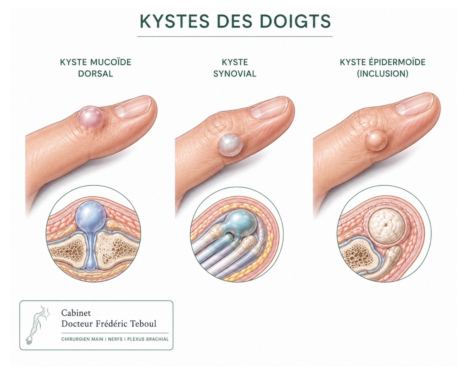
1. The synovial cyst — by far the most common
The synovial cyst, also called a ganglion, accounts for about 65% of all soft-tissue masses of the hand and wrist. Current Oncology 2023 It affects women about three times more often than men.
It is a pocket filled with thick, jelly-like fluid, arising from a joint or a tendon sheath, to which it often stays connected by a fine channel. One point specialists stress: it has no true cell wall, so strictly speaking it is not really a "cyst."
It should not be confused with a cyst of the interosseous scapholunate ligament — intra-articular, small, and painful when the wrist is extended (the "push-up" position) — which is also felt on the back of the wrist but right over the space between the scaphoid and the lunate.
In the finger, it tends to develop along the flexor tendon sheath, as a small firm bead that can get in the way of gripping. It is most often a cyst of the A1 or A2 pulley of the flexor tendon sheath. There it can be mistaken for the nodule of a trigger finger. See the trigger finger article.
Other common cysts
The carpal boss: a disorder of the joint between the carpal bones and the metacarpals. A very common condition, most often revealed by a dorsal cyst over the painful area.
The dermoid cyst: benign, it is neither a synovial cyst nor a pulley cyst. It is a pseudo-fluid cyst fed by sebum from a hair follicle trapped under the skin of the hand after a minor injury. A history of a pinpoint wound a few months or years earlier points to this diagnosis. Treatment is surgical only. It is filled not with jelly-like synovial fluid but with sebum.
What to know: many resolve on their own, and simple monitoring is reasonable when they cause no trouble.
In short, when faced with a dorsal synovial cyst of the wrist, one must be certain it is isolated. In some cases it can point to underlying conditions (involvement of the carpal bones, wrist osteoarthritis, arthritis, carpal boss, and others) for which specific treatments must be considered. In more than 90% of cases, however, the synovial cyst is strictly benign and spontaneous, with no underlying cause.
2. The mucous pseudocyst — the one at the fingertip
A small translucent or flesh-colored cyst on the back of the finger, between the last joint and the base of the nail.
Its distinctive feature is that it almost never occurs alone: it is closely tied to osteoarthritis of that joint and the bony outgrowths that go with it. That is why it appears mostly after age 40, and treating it without addressing the underlying joint invites it to return. See the finger osteoarthritis article.
It can press on the nail matrix and cause a groove or ridges in the nail (nail dystrophy) — a sign that often worries people more than the cyst itself, and that usually resolves once the lesion is treated.
3. Giant cell tumor of the tendon sheath — the second most common
This is the second most common mass in the hand, after the synovial cyst. It presents as a firm, slow-growing, often painless mass developing against a flexor tendon sheath.
The issue is not its nature — it is benign — but the quality of its removal. Recurrence rates reported in the literature vary and are sometimes high (10 to 30% of cases), especially when there are satellite nodules, a nearby joint, or a bony imprint visible on the X-ray.
In practice: it is a lesion that requires complete removal, under magnification, exploring its entire course. That is what makes the difference between a satisfactory operation and a repeat procedure two years later.
4. The glomus tumor — tiny, very painful, and long overlooked
This one is worth dwelling on, because it is the best illustration of what a specialist's opinion can change.
It is a small benign tumor arising from a microscopic structure involved in temperature regulation, very abundant at the fingertips. It often measures a few millimeters and most often sits under the nail. It is sometimes located in the finger pad and can, very rarely, be present on several fingers.
The three signs that, together, should raise suspicion
—Intense pain, out of all proportion to the size of the lesion, often coming in flare-ups.
—An extremely precise painful spot: pressing a point on just a few millimeters triggers the pain, while the rest of the finger is painless.
—Pain triggered by cold.
A fourth element is very suggestive: anti-inflammatories have no effect. If you have been taking them for months with no benefit at all, that is one more argument.
The average delay between the first symptoms and diagnosis is about seven years. Cases diagnosed after fifteen, or even forty, years have been published. The reason is simple: the lesion is rare, tiny, invisible, and the symptoms are readily blamed on something else — a nail infection, Raynaud's disease, a psychological complaint, a bone tumor, a trigger finger. Glomus tumor
The outcome, though, is very favorable: complete removal of the lesion eliminates the pain, and recurrences are rare when the excision is complete. In other words, years of suffering can end after the removal of a tumor a few millimeters across.
And the others
The lipoma
A soft, mobile, painless mass made of fatty tissue. Rare in the finger, it can compress a nerve when it develops near a nerve's path.
The enchondroma
A benign cartilage tumor developing within the bone of a phalanx, often found by chance on an X-ray, sometimes when a fracture occurs after a minor injury.
The nodules of Dupuytren's disease
These are not tumors as such, but a thickening of the fibrous membrane of the palm. They are frequently mistaken for a mass.
Nerve tumors, or schwannomas
Located within the nerves of the hand.
Vascular tumors
Arteriovenous malformations, hemangiomas, arteriovenous fistulas.
Synovial sarcomas
A malignant tumor of the hand that presents like a benign one.
And many others, rarer still.
How the diagnosis is made
The approach is methodical, and it begins with no test at all: how long the mass has been there, how fast it is growing, whether or not it is painful, its consistency, and how it moves relative to the skin and the deep tissues.
The role of ultrasound
It is the first-line test in this situation, for a very practical reason: it immediately answers the most useful question — is this a pocket of fluid or a solid mass?
A synovial cyst, superficial and fluid-filled, is easily seen on ultrasound. The test also clarifies how the lesion relates to the tendons and joints, and can guide an aspiration.
X-ray looks for involvement of the bone. MRI is reserved for deep, atypical, or suspicious lesions. In case of genuine doubt, a biopsy is done before any removal, in a specialized setting.
What to do, depending on the situation
A point often misunderstood: choosing aspiration over surgery is not a bad choice, provided you know the lesion may come back. Many patients prefer a simple procedure, even if it has to be repeated, over an operation. That reasoning is perfectly sound — it only requires having the information beforehand, not afterward.
Malignant lesions: rare, and not to be ignored
They exist, they are uncommon in the hand, and they are managed in centers specialized in musculoskeletal oncology.
Two principles capture the essentials:
A suspicious lesion is not operated on like a benign one. A removal done without a prior diagnosis can complicate later care. That is why biopsy precedes excision when there is doubt.
The warning signs listed at the start of this article are not there to make a diagnosis. They are there to tell you when to seek an opinion rather than wait.
It should be said plainly: fear of cancer is the leading reason for seeking care for a lump on the hand, and it is rarely justified. But it is a perfectly legitimate reason, and a visit for nothing is better than a mass watched for two years without ever being examined.
Frequently asked questions
Glossary
A pocket filled with jelly-like fluid, arising from a joint or a tendon sheath. The most common mass in the hand.
A small cyst on the back of the finger, near the nail, associated with osteoarthritis of the last joint.
The second most common benign mass in the hand, developing against a tendon.
A small, very painful benign tumor, often under the nail, arising from a temperature-regulating structure.
A benign cartilage tumor developing inside a bone of the hand.
Complete surgical removal of a lesion.
References
Soft Tissue Masses of the Hand: A Review of Clinical Presentation and Imaging Features. Current Oncology. 2023 — synovial cysts account for about 65% of masses of the hand and wrist; most lesions are benign
Common Soft Tissue Tumors Involving the Hand with Histopathological Correlation. PMC6702939 — the synovial cyst, superficial and fluid-filled, is easily seen on ultrasound
Giant Cell Tumor of Tendon Sheath — giant cell tumor of the tendon sheath is the second most common tumor in the hand, after the synovial cyst
Glomus tumor: a rare differential diagnosis for subungual lesions. 2024. PMC11437606 — characteristic triad, average diagnostic delay of about seven years, case reported at forty years
Glomus Tumors: Symptom Variations and Magnetic Resonance Imaging for Diagnosis. PMC3724001 — symptoms are frequently attributed to a chronic nail infection or a trigger finger
Delayed diagnosis and surgical management of a subungual glomus tumor. 2024 — reported average delay of seven years and four months between symptom onset and diagnosis
Article expanded and reviewed by the Dr Frédéric Teboul, surgeon of the hand, brachial plexus, and peripheral nerves, member of the Académie Nationale de Chirurgie.
Last updated: August 20, 2026
Disclaimer. This article is for information only. It does not replace a medical consultation. Only a clinical examination can establish a diagnosis and choose a treatment suited to your situation.
Author of this publication
Every piece of content is signed by its author and reviewed before publication.
 Author
Dr Frédéric Teboul
Hand surgeon — specialist in the brachial plexus and peripheral nerves
Author
Dr Frédéric Teboul
Hand surgeon — specialist in the brachial plexus and peripheral nerves