
Wrist pain that has been present for months, without any notable injury, in a young and active person. An X-ray showing one small bone that is "whiter" than the others, or an MRI that speaks of necrosis. And an unfamiliar, slightly worrying word: Kienböck.
This rare disease has a feature that makes it puzzling, for patients and doctors alike: what is seen on the images and what the patient feels do not always match. This article explains what it is, what we really know about it — and what we still don't.
What is Kienböck's disease?
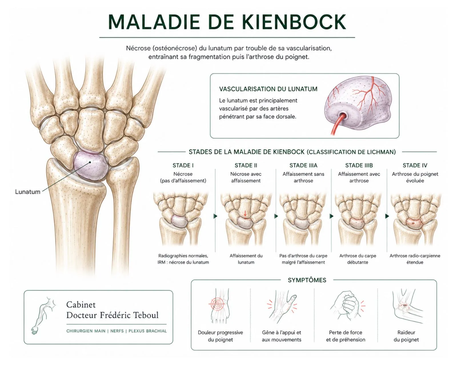
At the center of the wrist sits a small crescent-shaped bone, the lunate — doctors also call it the lunatum. It holds a key position: it links the forearm to the rest of the wrist bones and transmits a large share of the forces that cross the joint.
In Kienböck's disease, this bone loses part of its blood supply. This is called osteonecrosis, or avascular necrosis: without enough blood flow, the bone tissue weakens, may crack, collapse, and then fragment.
What makes it unusual among wrist conditions. It is neither age-related wear, nor the aftermath of a single fracture, nor a tendinitis. It is a problem with the blood supply to a bone, and its cause remains largely unexplained. This has a direct consequence: you cannot always tell a patient why it happened, or how it will progress.
Who is affected, and why — the hypotheses
The disease most often affects young adults, between 20 and 40 years old. Its exact cause is unknown, but several contributing factors are proposed, though none explains every case:
An anatomical peculiarity in the length of the forearm bones: an ulna slightly shorter than the radius — a negative ulnar variance — is frequently found. It may change how load is distributed on the lunate.
Repeated microtrauma, linked to manual or vibrating activity, which may weaken the bone and its blood supply.
Certain systemic diseases affecting bone circulation, more rarely involved.
To state clearly to the patient: these factors are associations, not certainties. In most cases, no single cause can be identified, and nothing the person did on its own explains the disease.
The symptoms
The signs set in gradually, often without any clear triggering injury:
Pain in the center of the wrist, on the back of the joint, at first with effort and then more constant.
Stiffness and a gradual loss of mobility, especially in extension.
A loss of grip strength, difficulty gripping or bearing weight.
Slight swelling on the back of the wrist.
These symptoms are not very specific: they resemble those of many other wrist conditions, which is why the diagnosis is often made late, sometimes after months of progression.
The most puzzling aspect of this disease
What the imaging shows and what the patient feels do not always go together. A lunate that looks severely damaged on X-rays may cause little pain, and the reverse also happens.
This lack of correspondence has a major practical consequence: the decision to treat, and how, is never based on the image alone. The actual discomfort, how long it has lasted, the patient's age and needs matter as much as the radiographic stage.
Diagnosis and staging
The diagnosis combines the clinical examination and imaging:
It may be normal at the very beginning. Later, it shows a denser lunate — "whiter" — then collapse and deformity.
It is the key test for early forms: it detects the impaired blood supply before the X-ray shows anything at all. It is what allows an early diagnosis.
It clarifies the state of the bone, looks for a fracture or fragmentation, and helps pinpoint the stage precisely.
The progression is classically described in stages — the Lichtman classification — ranging from involvement visible only on MRI to collapse of the lunate and osteoarthritis of the wrist. These stages guide the treatment discussion, but their boundaries are not always clear-cut, and on their own they are not enough to decide.
Treatment: an uncertainty that must be acknowledged
This is the point where we have to be honest, because it is what sets this disease apart from most others.
What the literature really says
No treatment has proven superior to the others. Systematic reviews conclude that the different techniques all provide pain relief and improved function, without any one standing out clearly.
The natural history is poorly understood, and it is not certain that surgery does better than non-surgical treatment in every situation. Some studies report stable, low-pain wrists in the long term without surgery.
This is not a reason to do nothing — it is a reason to decide case by case, with a hand surgeon, rather than applying a set recipe.
This uncertainty should not cause added worry: it simply invites caution toward websites that present a fixed, ready-made path. The general logic, for its part, is clear, even if the details are open to discussion.
At early stages: relieve pain and try to protect the bone
When the lunate has not yet collapsed, two approaches coexist.
Non-surgical treatment — rest, temporary immobilization, anti-inflammatories, activity modification — aims to ease the pain and rest the bone. It is often offered first, and is enough in some cases.
Early surgery, on the other hand, seeks to act on the presumed mechanism, in three ways:
When the ulna is too short, the radius can be shortened to rebalance the load and reduce the stress on the affected bone.
So-called revascularization techniques transfer a vascularized bone fragment to try to bring the lunate "back to life." They are suited to stages where the bone has not yet collapsed.
Other, simpler techniques aim to stimulate revascularization at a distance. Long-term series report good results on pain at early stages.
At advanced stages: treat the wrist, not just the bone
When the lunate has collapsed and the wrist begins to wear, the goal changes: it is no longer about saving the bone, but about relieving a damaged wrist.
The procedures then overlap with those for wrist osteoarthritis, chosen according to how far the wear has spread:
Partial wrist fusions, which join a few joints together while preserving some movement.
Removal of the first row of carpal bones (proximal row carpectomy), with or without an RCPI implant.
Removal of the lunate combined with a stabilizing procedure, in certain forms.
Total wrist fusion, reserved for very advanced and painful forms, as a last resort and after other treatments have failed on a stiff wrist.
Here again, comparisons do not point to any clearly superior technique: they relieve pain and improve function to a broadly similar degree, with differences in mobility depending on the procedure.
The overall picture
This table gives the logic, not a rule: at each step, several options are open to discussion, and the choice depends as much on the patient as on the image. That is one more reason to rely on a specialist's opinion rather than on a conclusion found online.
What can be said about the prognosis
Many patients keep, in the long term, a functional and low-pain wrist, with or without surgery, even if mobility and strength often remain slightly reduced. The outlook is therefore not one of inevitable worsening.
But the course is variable and hard to predict for any given individual, which justifies regular follow-up rather than a firm prognosis from the outset. The right stance is neither worry nor false reassurance: it is monitoring.
Recovery and follow-up
No general scheme can be given: specific, contextualized cases are presented in our knowledge center.
Frequently asked questions
Glossary
Small central bone of the wrist, crescent-shaped, affected in Kienböck's disease.
Death of bone tissue from a lack of blood supply; also called avascular necrosis.
An ulna slightly shorter than the radius; a factor frequently associated with the disease.
A description of the disease in stages, from the earliest (visible only on MRI) to the most advanced (collapse and osteoarthritis).
Surgical techniques aimed at bringing blood back to the lunate to try to revive it.
A procedure that changes the length of a forearm bone to unload the lunate.
References
Lichtman DM, et al. Kienböck disease: a new algorithm for the 21st century. Classification into stages I to IV — from involvement visible only on MRI to collapse and osteoarthritis
Lutsky K, Beredjiklian PK. Kienböck disease. J Hand Surg Am — poorly understood natural history, no correlation between image and symptoms, no strong evidence favoring any one treatment
Innes L, Strauch RJ. Systematic review of the treatment of Kienböck's disease in its early and late stages. J Hand Surg Am. 2010;35(5):713-717 — no significant difference between treatment groups for pain
Treatment of Stages IIIA and IIIB in Kienbock's Disease: A Systematic Review. PMC7708034 — all modalities provide relief; non-salvage procedures give results similar to salvage procedures
Charron BP, Chan KTK, et al. Systematic Review of non-salvage procedures in stage IV. 2024
Radius Core Decompression for Kienböck Disease Stage IIIA: Outcomes at 13 Years Follow-Up. J Hand Surg — good long-term results on pain at early stages
The Natural History of Kienböck's Disease Diagnosed at a late age. CiOS — in patients followed for an average of 7.5 years without surgery, stable radiographic indices and no osteoarthritis
Epidemiology and distribution by age and sex
Association between negative ulnar variance and Kienböck's disease
Read and reviewed by Dr Frédéric Teboul, surgeon of the hand, brachial plexus and peripheral nerves, member of the Académie Nationale de Chirurgie.
Last updated: August 20, 2026
Disclaimer. This article is intended for information only. It does not replace a medical consultation. Wrist pain that persists without an obvious cause deserves a specialist's opinion.
Author of this publication
Every piece of content is signed by its author and reviewed before publication.
 Author
Dr Frédéric Teboul
Hand surgeon — specialist in the brachial plexus and peripheral nerves
Author
Dr Frédéric Teboul
Hand surgeon — specialist in the brachial plexus and peripheral nerves